Sinus Pathology Imaging
April 1, 2018
April 1, 2018


PANO and CT Images taken on PaX-i 3D Green and
Periapical image was taken with the Vatech EzP Sensor.
INTRODUCTION:
- 52 year old male
- Presented with chief complaint of a pimple in the maxillary right quadrant.
- After an oral evaluation we took a panoramic image as a overview.
- During the patient interview and history, the patient expressed a strong desire to try to save the teeth in that area at all cost.
PATHOLOGY:

Looking at a magnified view, on the panoramic film, my initial diagnostic feelings were that the distal root of tooth #3 was creating the draining sinus tract.
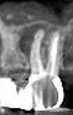
Following the ALARA Principle I chose to take a periapical radiograph for additional information and to get a clearer image of the area so as to confirm my diagnosis.

The periapical radiograph seemed to confirm my original diagnosis but showed that the area of the periapical pathology extended over to the mesial buccal root of tooth #2. (Progressing) At this point I had concerns regarding the most predictable course of treatment for my patient. I chose, with patient consent to take a cone beam ct image of the area.
3D RADIOGRAPHS:
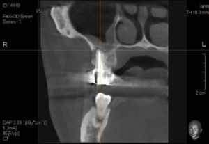
- Magnified view of the sinus perforation

- The cone beam image clearly shows that the periapical pathology is associated with the mesial buccal root of tooth #2.

- a coronal view of the mesial buccal root of tooth #2 shows a communication with the maxillary sinus and consequent pathology in the sinus.


- This volume rendering shows the loss of the buccal plate of bone on the mesial buccal root of tooth #2.
CONCLUSION:
The panoramic image and the cone beam images were taken by our PaX-i3D Green. The periapical image was taken with the Vatech EzP Sensor. Most importantly, this case shows how through the utilization of cone beam imaging we were able to make a definitive diagnosis and ultimately create a treatment plan with a more predictable outcome.
To discuss this case, please visit the Customer Portal.
Prosthetic Design and Fabrication of a Straumann...
As previously described, non-restorable teeth were removed and grafted. Following integration of the graft, 4 Straumann BLT implants were strategically placed using the Engel protocol. The implants were allowed to...

Cone Beam Computed Tomography in Implant Dentistry:...
In implant dentistry, three-dimensional (3D) imaging can be realised by dental cone beam computed tomography (CBCT), offering volumetric data on jaw bones and teeth with relatively low radiation doses and...

Periapical Radiograph Not Diagnostic – Power of...
Patient came in with pain, upper left posterior molar... a periapical was inconclusive... no swelling, no fistula. How can we determine the proper treatment plan today without CBCT..? An essential...

Not a Member yet? Sign up for Free
Get Started
 Protection